Cystoscopy
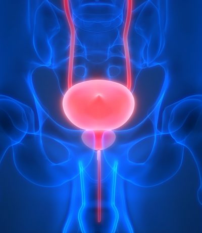
If the bladder needs to be examined in detail, a urologist may perform a cystoscopy.
A cystoscopy is a procedure that involves accessing the bladder from the tube that transports urine (urethra) with a long tube with an attached lens called a cystoscope. Used for both diagnostic and treatment purposes, a cystoscopy may be performed by a urologist as an outpatient procedure with or without sedation or with a local anesthetic.
- In some situations, the procedure is done in a hospital under general anesthesia.
- The specific way a cystoscopy is performed will depend on the reason why it’s being done.
Reasons to Have a Cystoscopy

For investigative purposes, a cystoscopy may be done to determine the reason why a patient is having recurring urinary tract infections or bladder infections. The procedure may also be done to diagnose bladder diseases, bladder inflammation (cystitis), or enlarged prostate (benign prostatic hyperplasia). A cystoscopy might also be done to treat certain bladder problems, such as ones involving small tumors, with special instruments.
Preparation
How It’s Done
An outpatient cystoscopy is usually done in a urologist’s office. Just prior to preparing to insert the scope, the bladder should be emptied. Patients lie down on a table, usually with feet in stirrups. Some type of sedation may be done, although this isn’t always necessary. Numbing jelly makes it easier to insert the flexible scope into the urethra.
If a cystoscopy is being done for treatment purposes, a larger scope is often used so instruments can be safely inserted. A larger scope is also used to take tissue samples.
After a Cystoscopy
Results may be discussed with a patient immediately after the procedure is completed. If a biopsy was taken, results won’t be known until lab tests are done. There may be some slight irritation following a cystoscopy. Staying hydrated and taking a warm bath may ease discomfort experienced following the procedure.
A cystoscopy is just one of several diagnostic procedures a urologist may perform to determine what’s causing a patient’s symptoms. Oftentimes, what’s learned from a visual inspection of the bladder and nearby structures is combined with results from urine and blood tests and ultrasounds and other image tests.
