KOELIS Trinity® Fusion Biopsy

illustrated-concept-of-cancer-detection

A well-developed treatment plan for prostate cancer is closely tied to early and accurate detection.

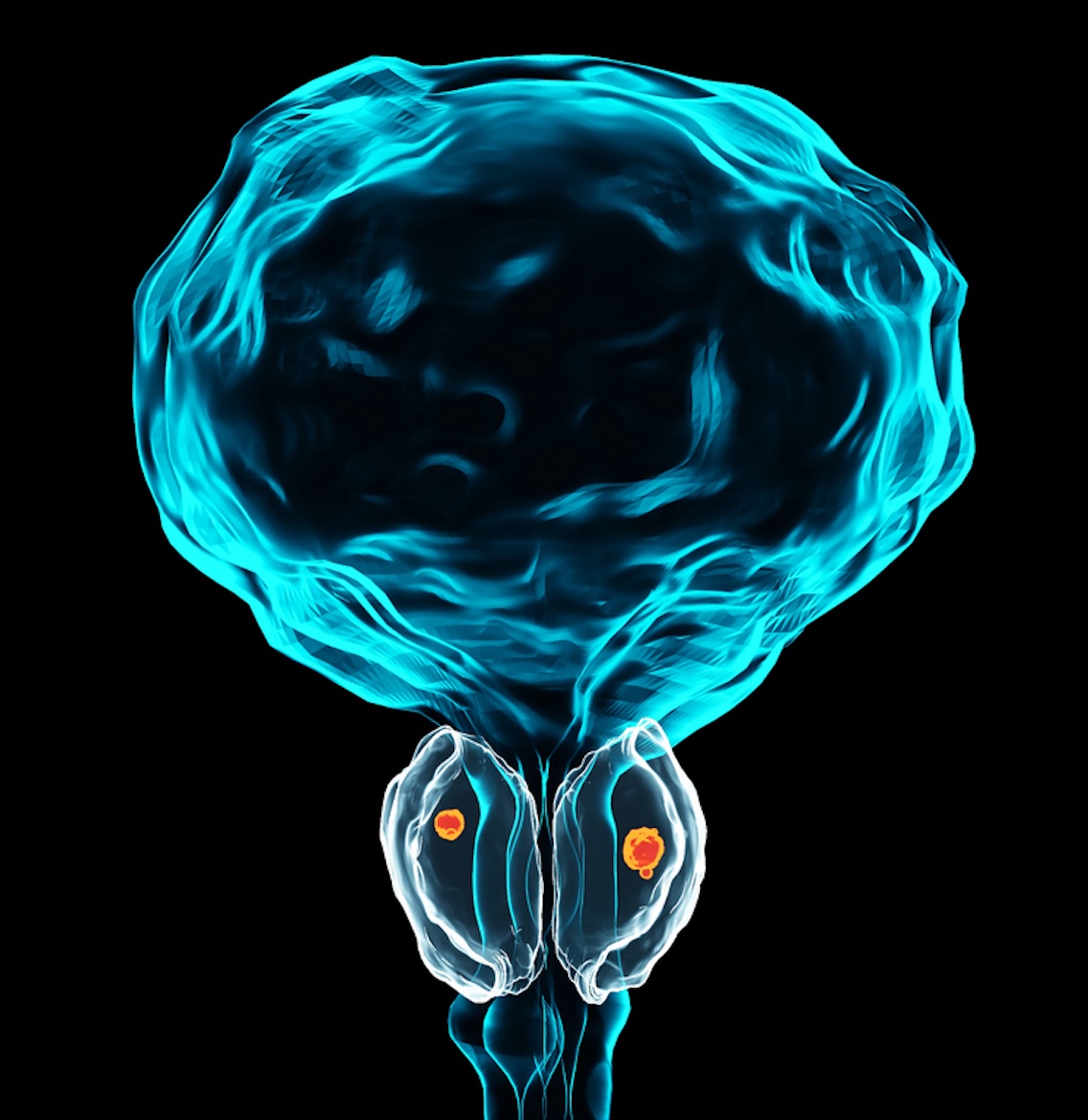
The prostate biopsy remains the most conclusive approach to prostate cancer diagnosis and involves the removal of small samples of prostate tissue to test for cancer presence, size, and aggressiveness.

  • A standard prostate biopsy performed via ultrasound imaging uses a random sampling technique.
  • The random sampling technique can lead to suspicious lesions being missed.

KOELIS Trinity® Fusion Biopsy Technology

MRI and ultrasound images are fused together
Targets suspicious areas in real time
Helps detect aggressive prostate tumors

KOELIS Trinity® is the most advanced MRI and ultrasound fusion system for targeted prostate biopsy. With the fusion approach, MRI images and ultrasound work together to detect aggressive tumors and help the physician to target specific areas in real time.

What Patients Can Expect

Your physician may perform a digital rectal exam or PSA blood test that suggests signs of prostate cancer and choose to confirm with a prostate biopsy.
1
Using the KOELIS Trinity® 3D technology, segmented MRI images are fused with the model generated with the ultrasound.
2
The fusion process results in a 3D model that serves as a map of your prostate.
3
The model provides a detailed representation of the prostate down to its exact size and shape.
Benefits of KOELIS Trinity®

The technology of the fusion biopsy helps physicians to detect potential cancer sites via the MRI before performing the biopsy.

The biopsy needle is precise within a few millimeters.
Every biopsy location is precisely mapped.
Biopsy locations are stored for future reference.
Stored data is invaluable during follow-ups.

Guiding Treatment Options

Detailed diagnosis helps personalize treatment planning.
Patients with low-risk cancer may choose active surveillance.
Stored exams help evaluate changes over time.
Fusion imaging supports more accurate treatment decisions.

The detailed diagnosis obtained through fusion biopsy helps your physician to personalize your treatment plan without over-treating you. For example, patients with low-risk prostate cancer can choose active surveillance to monitor cancer progression over time. Since the KOELIS Trinity® exams are stored for future reference in the system, the fusion approach can lead to higher accuracy in evaluating changes, leading to better treatment decisions.

Precision Biopsy and Follow-Up
1
Targeted Detection
With the fusion approach, MRI images and ultrasound work together to detect aggressive tumors and help the physician to target specific areas in real time.
2
Accurate Prostate Mapping
Using the KOELIS Trinity® 3D technology, segmented MRI images are fused with ultrasound models to create a detailed three-dimensional map of the prostate.
3
Long-Term Monitoring
Biopsy locations are precisely mapped and stored for future reference, allowing physicians to track changes and guide treatment decisions during follow-up evaluations.